Breast Cancer Diagnosis
Every year in Germany, approximately 70,000 women are diagnosed with a malignant tumor of the breast, meaning that about one in every eight to nine women will be affected by it during their lifetime. Breast cancer can also occur in men! In Germany, with about 500 new cases per year in men, it is significantly less common than breast cancer in women. If breast cancer is diagnosed, specialists from all disciplines are available to you at the Breast Cancer Center.
Focal points of treatment
Diagnostics
Three breast examinations are important:
- breast examination by touch (palpation)
- breast ultrasound (mammasonography)
- breast X-ray (mammography)
If these examinations reveal an abnormality in the breast tissue, it should be investigated further through a biopsy (tissue sample). In almost all cases, this can be done via a so-called core needle biopsy.
The clinical examination of the breast (and the armpits) consists of two parts:
Inspection
The doctor examines the breast to identify any changes in the skin or dimpling. The doctor will likely ask you to raise your arms, as some changes can only be detected this way.
Palpation
Palpation of the breast and armpits is used to detect changes in tissue (so-called lumps). If you feel a lump in your breast, this does not automatically mean you have cancer, as there are many benign lumps. A lump should always be further investigated (see Mammography/Mammasonography and Tissue Sample).
You should also perform these examinations regularly yourself. Ask your gynecologist to show you what to look for. Mammography and breast ultrasound are used to further investigate an abnormal palpation finding. These examinations can determine whether a lump is more likely to be benign or malignant. Another use for these examinations is in screening to detect potential tissue changes before they can be felt.
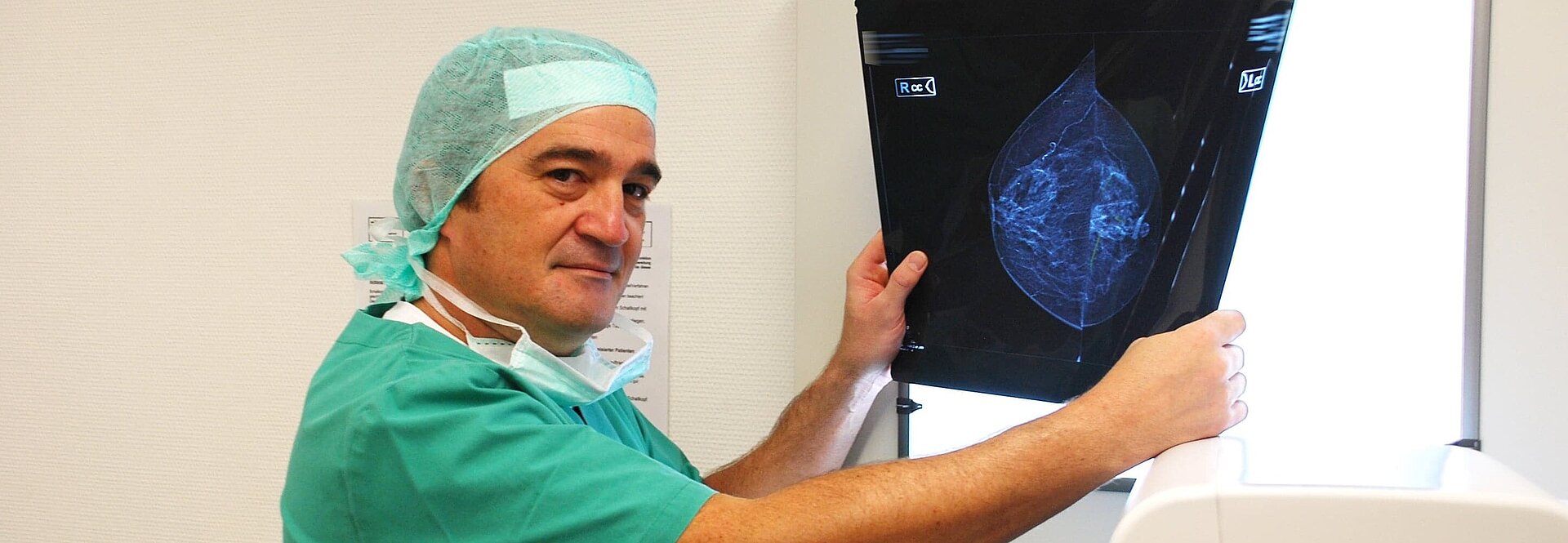
Mammography
Mammography is an X-ray of the breast. It can reveal changes in breast tissue. Even the smallest changes, such as so-called "microcalcifications," can be detected very early on. This allows the early stages of cancer to be identified and treated.
Breast ultrasound
Breast sonography is an ultrasound examination of the breast and the armpit. This procedure can also visualize changes in breast tissue as well as lymph nodes in the armpit. Especially in young women, breast tissue is very dense, making mammography difficult to interpret. In such cases, sonography is an important diagnostic method. Cysts in the breast can be clearly identified through sonography.
Surgical Treatment
Today, the majority of women with breast cancer can undergo breast-conserving surgery. During this procedure, the tumor itself, the immediate surrounding tissue, and some of the lymph nodes from the armpit are removed. Further analysis of the tumor tissue determines the likelihood of the disease recurring or metastases developing, and whether systemic therapy in the form of chemotherapy or hormone therapy is necessary.
In the case of breast-conserving surgery, follow-up radiation therapy is always required; this is less common after a mastectomy. Whether breast-conserving surgery is possible depends, among other things, on the location and size of the tumor in relation to the size of the breast. However, if a complete mastectomy is necessary, this by no means implies a loss of physical integrity, as the removed breast can be reconstructed. This breast reconstruction can be performed during the same surgical procedure—or at a later date.
In recent years, plastic surgery has developed various techniques that allow for the recreation of the breast shape without negatively impacting the patient’s disease progression. Reconstruction is performed either by inserting silicone gel implants or non-silicone-filled prostheses, or using the patient’s own muscle and skin tissue.
For some time now, new surgical techniques have been used that allow for the removal of lymph nodes in an even gentler and more targeted manner. Scientific studies have been and are currently examining whether, in the future, only the first lymph nodes reached by the lymphatic flow from the breast—the so-called “sentinel lymph nodes”—need to be removed. They are marked using dye solutions or radioactive substances, which are injected into the area surrounding the tumor prior to surgery so that they can then be specifically removed during the procedure. Using imaging techniques, the lymph nodes marked in this way are removed during the same surgical session and examined for tumor cells. If no tumor cells are found, it should be possible in the future to avoid the standard removal of additional lymph nodes from the armpit.
Radiation Therapy
Radiationtherapy is an integral part of breast cancer treatment. It is used after breast-conserving surgery and, depending on the stage of the disease, after mastectomies to minimize the risk of local recurrence. Treatment is administered daily, usually on an outpatient basis, and extends over a total period of about five to six weeks. We collaborate with all radiation oncology clinics in the area, ensuring that this necessary treatment can be provided close to home.
The goal of treatment is to achieve high effectiveness while sparing healthy tissue as much as possible. Protecting the skin and achieving a good cosmetic outcome are key considerations. Radiation is delivered using a linear accelerator with so-called ultra-hard X-rays. Tailored to each patient’s individual body contours, an optimal dose distribution is calculated at the start of therapy based on physical and medical criteria.
Drug Therapy
Drug-based tumor therapy encompasses all options for chemotherapy and hormone therapy. So-called high-dose chemotherapy is performed in collaboration with the Department of Internal Medicine and Polyclinic (Tumor Research). In addition, medications are used that aim to improve the quality of life for patients with advanced tumor disease who are undergoing chemotherapy. Innovative immunological antibody therapies for the targeted activation of the body’s own tumor defense mechanisms are also employed. The decision regarding any necessary drug therapy is made during the interdisciplinary tumor conference.


Outpatient clinics & appointments
Clinic Hours
Breast Cancer Center (Outpatient Clinic)
Monday and Wednesday 8 a.m. to 3 p.m. or by appointment
To make an appointment, call
Phone: 06142 88-1316
Email: brustkrebszentrum@~@gp-ruesselsheim.de
Coordinator
Mario Vescia
Tel.
06142 88-1316
Fax
06142 88-1223
vescia@gp-ruesselsheim.de

Oncology guideline program
Patient Guidelines for Breast Cancer
More InformationS3 Guideline on Early Detection, Diagnosis, Treatment, and Follow-up Care for Breast Cancer
More information
Interdisciplinary cooperation
Radiology
More informationOncology
more informationRadiation
Therapy More InformationPsychological Support
See Patient Information under P